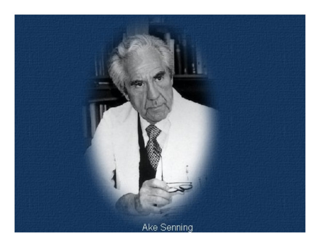
Ake SENNING
Célébrités en cardiologie
(Celebrities in cardiology)

Ake SENNING . © Heart and Coeur ©
 Ake SENNING
Réparation de Senning pour la transposition des grandes artéres
Ake Senning 1915-2000
On 21 July 2000, Ake Senning passed away in Zurich, at age 85, after a long illness. Dr. Senning's pioneering contributions to cardiovascular surgery will be an enduring legacy to humanity. He advanced almost every area of cardiac treatment, including open heart surgery, repair of congenital cardiac defects, pacemaker therapy, valve replacement, cardiac transplantation, and balloon angioplasty.
Born in 1915, in Sweden, Ake Senning attended medical school at the University of Uppsala and Stockholm. In 1948, he joined the innovative cardiovascular surgeon Clarence Crafoord, at Sabbatsberg Hospital in Stockholm. There, he helped develop one of the first pump oxygenators for cardiopulmonary bypass, which was successfully used in dogs in 1951. In 1953, this oxygenator was used to extract a left atrial myxoma; the patient”at that time, a young woman”is still alive today. Dr. Senning also promoted the use of hypothermia and cardioplegia and was the first to use elective fibrillation in heart surgery. In 1956, Dr. Senning became Associate Professor of Experimental Surgery at the University Thoracic Clinic of Karolinska Hospital, in Stockholm. Two years later, he introduced an atrial inversion operation (the Senning repair) for transposition of the great arteries (TGA). In this procedure, the atrial walls were reconstructed with flaps of autogenous tissue, which formed venous and arterial conduits; caval blood was rerouted to the pulmonary artery, while pulmonary venous flow drained into the aorta. This operation dramatically improved the prognosis for children with TGA. The Senning repair was superseded by the Mustard procedure in 1964 but later attracted renewed interest because of its effectiveness. Although the arterial switch procedure is now the preferred approach for treating TGA, the Senning operation remains a satisfactory alternative. On 8 October 1958, Dr. Senning made another major breakthrough when he placed the first implantable pacemaker in a 43-year-old man with Stokes-Adams syndrome. The pacemaker, designed by Rune Elmquist (of the Elema Company, now owned by St. Jude Medical), used two transistors and was about the size of a hockey puck. Six hours postoperatively, the device failed and had to be replaced by a second, identical model, which worked for 6 weeks. Forty years (and 26 pacemakers) later, the patient was enjoying a normal life at age 83.
In 1958, Dr. Senning also introduced autogenous fascia lata trileaflet valves for the replacement of diseased human aortic valves. These bioprostheses were later modified by other researchers and implanted in hundreds of patients. In 1961, Dr. Senning moved to Zurich to become Professor of Surgery and Director of the Surgical Clinic A at the University Hospital, where he remained until his retirement in 1985. There, he and his team performed the first heart transplant in Switzerland in 1969. When interviewed about this event, he stated that transplantation is technically simple. Und wenn man weiss, ist es kein Problem. (One must merely sew. And when one knows where to sew, there is no problem.)
After the advent of coronary artery bypass surgery in 1967, Dr. Senning helped to promote this operation in Europe. At the University Hospital, he was a colleague of Andreas Gruntzig, who introduced percutaneous transluminal coronary angioplasty clinically in 1977. Dr. Senning provided surgical standby during the first percutaneous transluminal coronary angioplasty procedures and coauthored the early reports about this technique. The recipient of countless awards and honors, Dr. Senning belonged to various professional surgical societies and was author or coauthor of at least 350 scientific publications. I knew Ake Senning personally, enjoyed his friendship, and admired his spirit. At is passing, the cardiovascular surgery profession lost a great pioneer and fine human being. He is survived by his wife Ulla and his 4 children (3 sons and a daughter). On behalf of the Texas Heart Institute, I salute Dr. Senning's memory and offer my sincere condolences to his family and colleagues.
 Ake SENNING
The pulmonary autograft procedure
The Ross Procedure, also known as the pulmonary autograft procedure for aortic valve disease, is a special surgical technique for replacing a diseased aortic valve.
The procedure was developed by the British surgeon Dr. Donald Ross in 1967.
Unlike other cardiac operations that use a mechanical valve or a tissue valve taken from an animal, the Ross Procedure replaces a patient's damaged aortic valve with his or her own pulmonic valve.
The pulmonic valve is used because it is the patient's own tissue and is identical in shape, size and strength to the aortic valve. Overall, the success rate for the Ross Procedure is over 97 percent and the long-term results have been excellent.
The Ross Procedure is designed for people who have significant aortic valve disease€”either stenosis or regurgitation or both. In the advanced stages, aortic valve disease can cause chest pain (angina), fainting during exercise, enlarged heart, shortness of breath or congestive heart failure.
Generally, valve replacement is when there are signs and symptoms of a progressive strain on the left ventricle, the main pumping chamber of the heart.
The left ventricle may become dilated and the wall thickened with extra muscle (hypertrophy). Symptoms may include heart pounding with exercise, reduced exercise tolerance or episodes of severe shortness of breath. In some patients, fainting, especially following exercise, may occur.
A variety of conditions may eventually lead to an abnormal aortic valve which requires replacement. A congenital condition, known as bicuspid aortic valve (the valve normally has three cusps), may over several decades lead to progressive leakage or serious obstruction. Rheumatic fever may involve the aortic valve. In certain families, a genetic trait causes weakness of the aortic valve and the wall of the aorta, producing a progressive leakage. In some older patients, the valve leaflets gradually become stiffened, heavily calcified and produce an obstructive condition known as calcific aortic stenosis. Aortic stenosis may also occur on a congenital basis.
No blood thinners (anticoagulants) are needed. If the aortic valve is replaced with a mechanical valve, the patient must take anticoagulants for the rest of his or her life. These medications have a small risk of excess bleeding or hemorrhage and the effects of the anticoagulants must be monitored with a blood test every 3 or 4 weeks and the dosage adjusted.
No postoperative deterioration by calcification of the valve—This can be a problem for patients who have their valve replaced with one from an animal (pig or cow). The younger the patient, the less durable the animal valve. The patient's pulmonic valve is the right size to replace the aortic valve.
No artificial material is used for the new aortic valve—This avoids many problems including rejection. Since the new aortic valve is created from the patient's own tissue, the tissue is alive and healthy, instead of being frozen or chemically treated.


